Amir Shakerian1,2 ![]() ,
Permal Deo1,
Ebrahim Rahimi2,
Ali-Reza Shahjavan3,
Faham Khamesipour4
,
Permal Deo1,
Ebrahim Rahimi2,
Ali-Reza Shahjavan3,
Faham Khamesipour4
For correspondence:- Amir Shakerian Email: amshakerian@iaushk.ac.ir Tel:+983833361045
Received: 28 January 2016 Accepted: 12 April 2016 Published: 27 May 2016
Citation: Shakerian A, Deo P, Rahimi E, Shahjavan A, Khamesipour F. Molecular detection of Brucella melitensis in sheep and goat milk in Iran. Trop J Pharm Res 2016; 15(5):913-918 doi: 10.4314/tjpr.v15i5.3
© 2016 The authors.
This is an Open Access article that uses a funding model which does not charge readers or their institutions for access and distributed under the terms of the Creative Commons Attribution License (http://creativecommons.org/licenses/by/4.0) and the Budapest Open Access Initiative (http://www.budapestopenaccessinitiative.org/read), which permit unrestricted use, distribution, and reproduction in any medium, provided the original work is properly credited..
Purpose: To detect Brucella melitensis in the milk of reared sheep and goats from Isfahan and Shahrekord regions, Iran.
Methods: A total of 225 milk samples (sheep = 125; goat = 100) were collected from Isfahan and Shahrekord regions, Central Iran. Polymerase chain reaction (PCR) was used to detect the presence of B. melitensis in the milk following standard procedures.
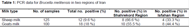
Results: From 225 milk samples, 20 (8.9 %) were positive for B. melitensis. Out of 125 sheep milk, 12 (9.6 %) had B. melitensis, and of these, 8 (66.6 %) were milk collected from Shahrekord and 4 (33.3 %) from Isfahan region. On the other hand, out of 100 goat milk samples, 18 (18 %) were positive for B. melitensis, out of which 10 (55.5 %) were from Shahrekord and 8 (44.4 %) from Isfahan.
Conclusion: The findings show that B. melitensis is present in a significant proportion of caprine and ovine milk in a section of Iran.
Introduction
Brucellosis in sheep and goats is caused by Brucella melitensis, one of the most virulent species of Brucella [1-2]. Brucellosis is a widespread zoonosis, especially in the Mediterranean and Middle-East regions of the world where it constitutes occupational and public hazard [3-6]. Brucella melitensis is transmitted via ingestion of contaminated meat and milk and contact with contaminated individuals or formites. Transmission of B. melitensis among caprine and ovine herds is rapid, and the disease being systemic in nature affects many organs and tissues [1,3,7]. Once the acute period elapses, symptoms of brucellosis becomes non-pathognomonic, and the incriminating organism then becomes chronically located in both supramammary lymph nodes and mammary glands of 80 % of infected animals [8].
Interest have been aroused to keep brucellosis under control in endemic regions, because of the economic and health impact of brucellosis [9,10]. It is necessary to identify new endemic regions and to implement strict eradication programs beyond national borders. There is increasing reports on the incidence of brucellosis [8,9]. Studies have revealed that in a few years to come, there is likely to be geometric increase in the incidence of brucellosis [8,9].
Diagnosis of brucellosis using fluid samples (such as milk and blood samples) is the cornerstone for the control and eradication of Brucella infection [11]. Traditional diagnostic methods such as culturing and serological tests (such as milk ring test) are often used for diagnosis of brucellosis. Culturing of Brucella species is difficult due to its fastidious nature and coupled with laborious biochemical tests for its identification [12]. The serologic method only detects the presence of Brucella antibody in serum of infected individual, it often gives false positive/negative reactions, cross-reactions between Brucella antigen is common including cross-reactions with other bacteria including Yersinia enterocolitica, Campylobacter fetus, Vibrio cholera, Bordetella bronchiseptica and Salmonella species [13,14], it is not specific, and does not distinguish active and non-active infection following post-treatment antibody responses [7,15,16]. Thus, these methods do not detect any Brucella species. Further, these methods are limited because they are time-consuming, low sensitivity when there is low amount of living Brucella organisms in the blood and the risk of laboratory personnel being infected following inhalation of aerosol droplets [13,17]. This makes treatment of the infection difficult and moreso limits efforts being made to reduce the prevalence of Brucella melitensis infection.
However, molecular detection using polymerase chain reaction (PCR)-based test has proved to be fast (> 4 h) and sensitive for the diagnosis of brucellosis [18,19]. Another advantage is that PCR is capabale of detecting few Brucella cells or the minutest Brucella gene copies in samples [20]. The aim of this study was to detect molecularly B. melitensis in the milk of sheep and goats reared in Iran.
Methods
Milk samples
This study was a cross- sectional conducted. A total of 225 apparently healthy animals (125 sheep and 100 goats) were sampled randomly. Milk samples consisting of 125 samples from Lori- Bakhtiary sheep breeds and 100 samples from traditional goat breeds, were collected in Isfahan and Shahrekord areas in central Iran. Sampled animals were members of flocks with a history of abortion. The milk samples were collected aseptically in a sterile cup with a lid and aseptically transported to the Islamic Azad University of Shahrekord laboratory within 10 minutes of collection. Samples were divided into 0.5 mL of sterile 2-mL Eppendorf tubes, and kept frozen (−20 °C) until used [7].
DNA extraction and PCR
Brucella melitensis DNA was extracted from milk by the method of Leal-Klevazas et al [20]. Briefly, frozen milk samples were thawed at room temperature and 400 μL of lysis solution (100 mM Tris-HCl (pH 8), 100 mM NaCl, 1 % Sodium dodecyl sulfate (SDS), 2 % Triton-x100) and 10 μL of proteinase K (10 mg/mL) were added to 400 μL of the fatty top layer of each milk sample. The contents were incubated at 50 °C for 30 min. Thereafter, 400 μL of saturated phenol (liquid phenol containing 0.1 % 8-hydroxyquinoline, saturated and stabilized with 10 mM Tris-HCl (pH 8) and 0.2 % of 2-mercaptoethanol) were added, mixed thoroughly and centrifuged at 8000 ×g for 5 min. The aqueous layer was transferred to a fresh tube and an equal volume of chloroform: isoamyl alcohol (24:1) was added, mixed thoroughly and centrifuged as described above.
The upper layer was again transferred to a fresh tube and an equal volume of 7.5 M ammonium acetate was added and mixed thoroughly. The contents were kept on ice for 5 min, centrifuged at 8,000 x g for 5 min and the aqueous content was transferred to a fresh tube. Two volumes of 95 % ethanol were added, mixed and the tubes were stored at –20 °C for 12 h. DNA was recovered by the final centrifugation as described above, the pellets were rinsed with 1 ml of 70 % ethanol, dried and resuspended in 30 μL TE buffer (Appli Chem, Darmstadt, Germany). In addition, a commercial DNA extraction kit (Dneasy® Tissue Kit, Qiagen, Hilden, Germany) was also used in the study. For this purpose, 25 μg of the fatty top layer were used as the initial extraction material. Subsequent extraction stages were applied according to the manufacturer’s recommendations. Extracted DNA was stored at -20 °C until processed.
Synthetic oligonucleotide design
The B. melitensis-specific primers used were previously described by Bricker and Halling [21]. The sequences of the primers were 5’-AAATCGCGTCCTTGCTGGTCT GA-3’ (B. melitensis-specific primer) and 5’-TGCCGATCACTTAAGGGCCTTCAT-3’ (IS711-specific primer from Sina Gene, Iran).
DNA amplification and detection of PCR products
PCR was carried out in a total volume of 50 μl, using 10 mM Tris-HCl (pH 9), 3 mM MgCl2, 50 mM KCl, 0.1 % Triton-x100, 200 mM of each of the four deoxynucleotide triphosphates (Lavora, Tellow, Germany), 0.4 mM of each primer (50 pmol), 2 IU of Taq polymerase (Fermentas, Opelstrasse 9, Leon-Rot, Germany) and 2 μl template. The amplification was performed in a DNA thermal cycler (Thermo, Px2 Thermal Cycler, USA) as follows: initial denaturation step at 94 °C for 4 min, and 35 cycles of 94 °C for 1 min, 60 °C for 1 min and 72 °C for 1 min. The final incubation was at 72 °C for 5 min [20,21].
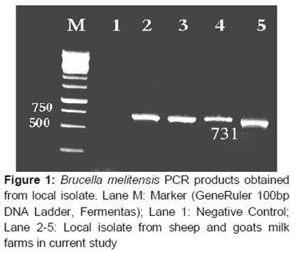
Amplification products were resolved in a 1.5 % (w/v) agarose gel containing 1xTBE buffer (100 mM Tris-HCl (pH 8), 90 mM boric acid and 1 mM Na2EDTA) and stained with ethidium bromide (0.5 μg/mL) and evaluated by a computerized image analysis system (Spectronics Co., Gl-5000, England). A visible band of appropriate size (731 bp) was considered as a positive reaction for B. melitensis. A positive control (based on DNA from B. melitensis 16 M) and a negative control (DNases and RNases free water, AppliChem) were included in all the tests. To check the reliability of the results and to detect any external contamination, all samples were processed in duplicate.
Determination of detection limit of PCR for inoculated milk
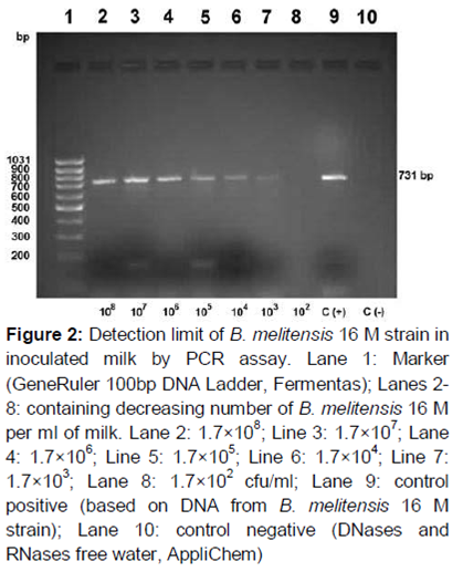
B. melitensis 16 M strain was grown on trypticase soy agar (TSA, Merck) at 37 °C for 48 h. A single colony was removed from TSA, placed in trypticase soy broth (Merck) and incubated at 37 °C for 48 h. Thereafter, the culture was prepared in sterile saline and 10-fold dilutions (from 10-1 to 10-10) were made. From these dilutions, 0.1 ml suspension was inoculated onto TSA plates and incubated at 37 °C for 48 h and the colonies present were then enumerated. The number of organisms in the dilutions was estimated spectro-photometrically at 623 nm and the concentration of the original B. melitensis culture was estimated as 1.7 × 10⁹ CFU/ml (OD 0.18). To assess the limit of detection of the PCR assays, 10 raw milk samples collected from Brucella-free sheep from the Research Farm of Faculty of Veterinary Medicine, Islamic Azad University Shahrekord Branch, Iran, were artificially contaminated with a known decreasing number of pure B. melitensis 16 M strain. The final concentrations of the organism in milk was 1.7 × 10⁸, 1.7 × 10⁷, 1.7 × 10⁶, 1.7 × 10⁵, 1.7 × 10⁴, 1.7 × 10³, 1.7 × 10² and 1.7 × 101 CFU/mL. B. melitensis DNA was extracted from all dilutions of milk, and processed by PCR as described earlier. The final concentrations of organisms were verified by plating onto TSA.
Statistical analysis
All statistical analyses were performed at 95 % confidence interval (CI) using Win Episcope version 2.0 programme. The level of significance was set at p < 0.05.
Results
Out of 225 milk samples, 20 (8.9 %) were positive for the PCR of B. melitensis. Out of 125 sheep milk, 12 (9.6 %) were positive for PCR of B. melitensis. Of these, 8 (66.6 %) were milk collected from Sharekhord while 4 (33.3 %) were from Isfahan region. Out of 100 goat milk, 18 (18 %) were positive for the PCR of B. melitensis. Of these 10 (55.5 %) were from Sharekhord while 8 (44.4 %) were from Isfahan ( and ).
No significant differences (p > 0.05) were found between sheep and goats milk samples positive for B. melitensis in Isfahan and Shahrekord, Iran. A positive PCR result on the ethidium bromide stained agarose gel was detected with different aliquots containing B. melitensis at a density of at least 1.7 × 10³- 1.7 × 10⁴ CFU/ml in milk ().
Discussion
PCR assay is a specific and sensitive choice for the detection of different bacterial agents [2,22,23]. In this study, 9.6 % (12/125) of ovine and 18 % (18/100) of caprine milk samples were positive for PCR of B. melitensis biovar 3. This suggests that sheep and goat infection by B. melitensis in Iran is relatively lower when compared with other regions. For instance, Erdenlig and Sen [24] reported 88.5 % B. melitensis biovar 3 among 78 Brucella isolates from different regions of Turkey [26]. In Central Anatolia region of Turkey, 94.8 % among 39 Brucella isolates from sheep were B. melitensis biovar 3 [25].
It has been reported that the detection limit of the PCR in milk samples range from 10 bacteria/ml [26], 1000 CFU/ml [11], 2.8 × 10⁴ CFU/ml [18] to 4.2 × 10⁴ CFU/ml [28]. In this study, 1.7 × 10³ - 1.7 × 10⁴ CFU/ml of B. melitensis 16 M strain was detected. The lower detection rate recorded in the current study may be attributed to the DNA extraction procedures.
The extraction procedure used in this study had successfully been employed in the detection of B. melitensis DNA in sheep/goat milk [26]. In a PCR study by Hamdy and Amin [27], 39 milk samples were collected from 21 sheep and 18 goats. In agreement with our results, Leal-Klevezas et al [20] also detected as positive a higher positive number of milk samples by PCR assay when compared to bacteriological culture methods [20].
The PCR results achieved in this study are in agreement with the results obtained in previous studies, i.e. less variable than the results of bacteriology or serology [3,20,24]. Deficient isolation techniques or the stage of infection may explain the superiority of the PCR assay to isolation methods. Moreover, PCR assay detects minutest traces of genetic material in samples, while culture methods detect only viable organisms [6].
The specificity of the primers used in the current study has been evaluated with a variety of microorganisms that have a close antigenic relationship with Brucella which causes false-positive results in serology, and the absence of amplification with DNA of these species has shown the primers to be specific for B. melitensis biovars 1, 2 and 3 [11,28]. In this study, B. melitensis DNA was detected in sheep and goat milk samples by PCR. Gupta et al [19] revealed that the sensitivity and specificity of PCR in detecting the presence of B. melitensis in goat milk were 90 and 100 %, respectively [19]; and our results showed almost the same sensitivity and specificity.
The determination of B. melitensis from sheep and goat milk samples is important because raw ovine/caprine milk is used in the production of traditional cheese in Isfahan and Shahrekord, Iran [6].
Conclusion
The findings of this study show that a sizeable percentage of sheep and goat reared in Iran are infected with B. melitensis biovar 3 which is excreted in their milk. Consumption of traditional cheese may be a route for transmission of brucellosis to humans. Control of brucellosis in animals will subsequently result in decreased incidence of the disease in humans. To effect rapid and accurate diagnosis of Brucella load/status of caprine/ovine milk for human consumption is paramount for public health.
Declarations
Acknowledgement
References
Archives


News Updates